
OPTOS Retinal Exam
Our office highly recommends Optomap retinal screenings because:
- It allows for an enlarged image of the retina revealing greater detail.
- It takes just a few minutes start-to-finish, rather than approximately 45 minutes with dilation.
- You leave the office with vision intact, rather than with light-sensitivity and blur for 4-6 hours (dilation).
- Creates a permanent record.
- Allows for future comparisons–we can compare this year’s image to next year’s image—side by side.
- Can be reviewed by other doctors, if necessary, for medical conditions or co-management with a surgeon.
Many eye problems can develop without you knowing. You may not even notice any change in your sight. But, diseases such as macular degeneration, glaucoma, retinal tears or detachments, and other health problems such as diabetes and high blood pressure can be seen with a thorough exam of the retina.
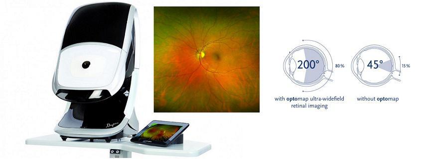
Optos
Annual eye exams are vital to maintaining your vision and overall health. We offer the optomap® Retinal Exam as an important part of our eye exams. The optomap® Retinal Exam produces an image that is as unique as you fingerprint and provides us with a wide view to look at the health of your retina. The retina is the part of your eye that captures the image of what you are looking at, similar to film in a camera.
Many eye problems can develop without you knowing. You may not even notice any change in your sight. But, diseases such as macular degeneration, glaucoma, retinal tears or detachments, and other health problems such as diabetes and high blood pressure can be seen with a thorough exam of the retina.
An optomap® Retinal Exam provides:
- A scan to show a healthy eye or detect disease.
- A view of the retina, giving your doctor a more detailed view than he/she can get by other means.
- The opportunity for you to view and discuss the optomap® image of your eye with your doctor at the time of your exam.
- A permanent record for your file, which allows us to view your images each year to look for changes.
The optomap® Retinal Exam is fast, easy, and comfortable for all ages. To have the exam, you simply look into the device one eye at a time and you will see a comfortable flash of light to let you know the image of your retina has been taken. The optomap® image is shown immediately on a computer screen so we can review it with you.
Please schedule your optomap® Retinal Exam today!
For more information on the optomap® Retinal Exam, go to the Optos website
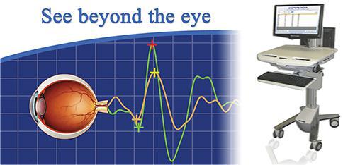
Visual Evoked Potential
VEP measures the electrical activity in the vision system. When light from an image enters your eye, it is converted into electrical energy at the retina and travels through the optic nerve to the visual cortex of the brain which processes vision. The Diopsys® NOVA-VEP test measures the strength of the signal reaching your visual cortex and how fast it gets there. The VEP technology in the Diopsys® NOVA device helps determine how your eyes communicate with your brain in a way that no other instrument or vision test can.
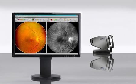
Cirrus OCT
The OCT allows for detection of other diseases such as macular holes, hypertensive retinopathy and even optic nerve damage. Using an OCT allows for early treatment in patients and dramatically improves the success of these treatments, especially in diseases such as wet macular degeneration – where the eye disease progresses rapidly.
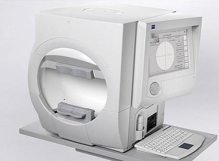
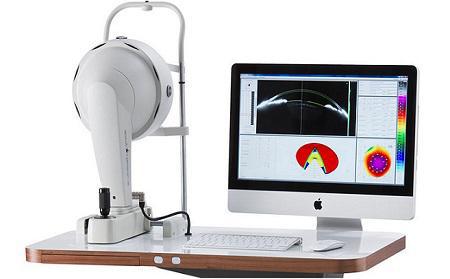
Humphrey Visual Field
The Field Analyzer is an important tool to detect and follow glaucoma and many other ocular conditions. Patients who either have glaucoma or are suspected of having glaucoma will undergo repeated testing with this instrument.
The procedure is performed quickly and easily in approximately 8-10 minutes. During the test, lights of varying intensities appear in different parts of the visual field while the patient’s eye is focused on a central spot. The results are compared against the normal healthy eye in order to determine if any damage has occurred.
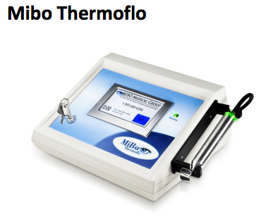
Our office offers a new and affordable solution for patients suffering from chronic dry eye. Mibo Thermoflo is a revolutionary dry eye treatment that uses continuous and controlled heat to the outer eyelids with a gentle massage. Ultrasound gel is used as a conductor in which the heat emitted from the device is then absorbed into the tissue and helps melt the meibum within clogged oil glands. This will maximize lubrication and help preserve tears offering instant relief to patients suffering from chronic dry eye.
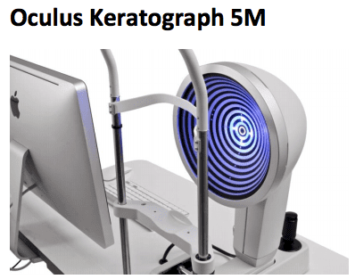
The OCULUS Keratograph 5M is a multifaceted instrument that combines many measurements and dry eye analysis suite into one device. The instrument uses Placido disc illumination to take measurements of the ocular surface, and different colors of light emitting diodes (LEDs) are used depending on the application. The Keratograph 5M offers Meibo-Scan for meibography of the upper and lower eyelid to check for Meibomian Gland Dysfunction (MGD), the most common cause of dry eyes, the TF-Scan for evaluation of the tear film break-up time, and the R-Scan for automatic bulbar redness classification. The Keratograph 5M features a high-definition color camera that can take both images and video. The device’s built-in software lets the doctor view 2D and 3D images as needed.